Nanotechnology may soon help brain surgeons navigate to the molecular level when treating brain tumors.
Medical researcher and nanotechnology professor Sam Gambhir and his research team have devised a way to use gold-based particles to image cancer cells in the brain. Although years away from practical use in humans, the research has the potential to advance the abilities of brain surgeons, who currently remove tumors while observing with the naked eye.
The nanoparticle the team is studying capitalizes on a tumor’s disruption of na

tural blood flow in the brain to collect around cancerous cells, enabling imaging of the areas.
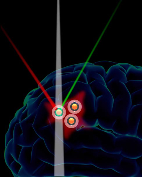
The imaging abilities developed by Gambhir are threefold. First, gold particles create a distinctive magnetic resonance imaging (MRI) signal that shows the precise tumor location in the brain. Second, a method called photoacoustic imaging exploits the sounds that gold emits when exposed to certain frequencies of light to create an image of the tumor. Last, a method called Raman spectroscopy employs a technique borrowed from counterfeit detection that monitors light emission spectra from the particle.
“Although their origins are in entirely different fields — in this case counterfeit detection — what we’re doing is taking these gold-core particles and making them work in a biological system,” Gambhir said.
These imaging methods could eventually serve the purpose of allowing surgeons to see tumors up close during surgery.
“Imagine that a surgeon eventually would have on molecular goggles, and those goggles let the surgeon see where the tumor is at the molecular level, thereby maximizing removal of just the tumor and sparing most of the healthy tissue,” Gambhir said.
The imaging could hold benefits both before and after surgery, as well. According to Gambhir, molecular-level awareness of cancer cells could also streamline treatment through more precise planning and more accurate evaluation of results.
Many obstacles still remain before these ideas can become a reality. The imaging has so far only been tested on human cancer cells growing in animals. Gambhir said that before the studies can move to a human brain, researchers would have to evaluate possible effects that gold particles could have on a person.
Gambhir noted that he is already working with the Food and Drug Administration to approve testing in a human bowel. He estimated that the first human pilot of a nanoscale-imaging procedure is around three years away, and if that pilot is successful, routine use could be around five years away.
“Why should surgery be limited to what the human eye sees?” Gambhir asked. “What you really should be seeing are microscopic molecular events.”
The study is significant not only for its potential medical applications, but also as collaboration between nanotechnology engineering and medicine, which are often disconnected fields.
“Work like this inevitably involves multiple disciplines, and so Stanford with its strength in both engineering and medicine … allows for technologies like this to appear and then eventually to be translated into humans,” Gambhir said.
This engineering precision is key in dealing with brain tumors because surgery requires leaving as many healthy cells behind as possible.
“In this work, what we showed was that by properly modifying these gold-based particles, we’re able to create three kinds of imaging signals that home very nicely and surprisingly to brain tumors of multiple types in animals,” Gambhir said. “They’re very effective at not getting into the normal brain.”