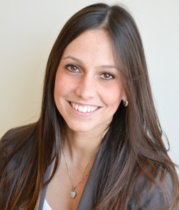
The research team—led by Livia Eberlin, a postdoctoral fellow in chemistry, and Richard Zare, chair of the Department of Chemistry and Eberlin’s advisor—employed mass spectrometry to more accurately assess the health of cells in surgically removed stomach tissue, presenting the potential for reduced surgery return rates.
Under current surgical procedures, the surgeon will remove tissue that he or she believes to be cancerous before sending the extracted sample to a pathologist. The pathologist then identifies the margins, or healthy edges, of the remaining tissue, albeit—due to the 20 to 30 percent chance of a diagnosis error—while potentially leaving some tumorous cells behind.
“Finding the margins is really important in surgery…[pathologists] want the margins to be clear of cancer,” Eberlin said. “[It] is really hard to say if there’s cancer there or not.”
By contrast, mass spectrometry involves turning molecules into charged particles and sending them down a vacuum tube. By measuring the amount of time it takes for each molecule to reach the end of the tube, the machine generates an image with thousands of peaks, each of which represents the abundance of a different chemical in the sample and thus reflects cell health.
In addition to Eberlin and Zare, the research team also included Assistant Professor of Surgery George Poultsides M.S. ’11 and Professor of Health Research and Policy Robert Tibshirani M.S. ’83 Ph.D. ’85.
Poultsides emphasized that all members of the team had been vital to the technique’s development, despite their disparate backgrounds.
“It was a risk,” Poultsides said. “It was a collaboration between researchers who do not speak the same language—a risk that paid off.”
Although most scientific research remains confined to a specific field, Poultsides framed the team’s output as reflective of the broader importance of bridging disciplines, particularly between clinicians and researchers.
“Clinicians and researchers are in their own silo,” Poultsides said. “It’s hard to move between disciplines, but Stanford is an institution where collaboration is encouraged. I felt it was very easy to work with chemists and a statistician.”
Even so, the team initially struggled to identify funding for the study, according to Poultsides. They eventually won support from the Stanford Hospital and Clinics Cancer Innovation Fund.
Team members said that they hope to introduce their instrument to pathologists on a clinical level in around a decade. In the interim, they will work to make the product more user-friendly while also testing it on a larger sample of cancer patients to refine its accuracy.
Contact Christina Gibbs at clgibbs ‘at’ stanford ‘dot’ edu.
