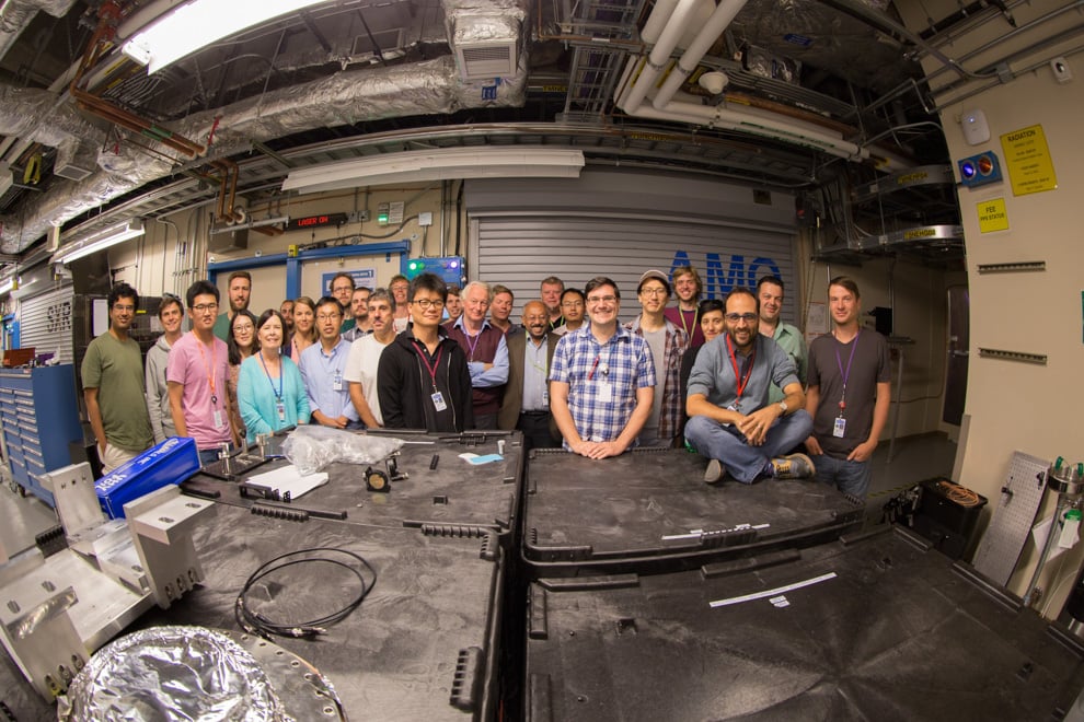
In a collaboration with other institutions, researchers from Stanford Linear Accelerator Laboratory (SLAC) have created the first 3D movie of a virus attacking a healthy cell. The movie is part of an initiative to use algorithms to visualize infections.
Of the 13 authors on the paper about the movie, which was published in “Nature Methods” last month, two of the researchers — Chun Hong Yoon and Andrew Aquila — were SLAC scientists.
Scientists used a highly sensitive x-ray laser, the Linac Coherent Light Source (LCLS), to take snapshots of the virus. Aquila called the LCLS, which is housed in SLAC, the “crown jewel of the national lab.” SLAC has the world’s largest x-ray facility.
The LCLS took snapshots of the PR772 virus in different orientations. The PR772 virus is relatively large, making it ideal for imaging. The snapshots left x-ray diffraction pattern — imprints that researchers interpreted using a new generation of algorithms designed for these extremely precise x-ray pulses. The process, called “manifold based analysis,” reconstructed 3D images of the viruses.
“In physics and biology, typically scientists build a large, bulky instrument like a spectrometer to make predictions; however, these machines can be expensive,” said lead researcher and University of Wisconsin-Milwaukee professor Abbas Ourmazd. “The cheapest way to make predictions is by using the brain; we can extract information using clever algorithms that our own brains devise. I hope this kind of algorithmically driven science is where the future of science lies.”
Viruses are known to attack host cells by altering their genetic makeup. The scientists’ movie gave further insight into the process, demonstrating how the virus under observation simultaneously rearranged its genetic material and formed a tubular structure to dispense its DNA into the host.
This simultaneous reaction documented by the scientists sheds light on viral mechanisms, particularly because previous methods of detecting biological structure relied on less sensitive predictions. One such method is cryo-electron microscopy, or cryo-EM, which transmits electrons to study samples at extremely low temperatures. Cryo-EM takes snapshots of biological samples but does not use mathematical analysis to morph them into a continuous, 3D movie.
Ultimately, the researchers want to make functional movies of the tiny structures responsible for keeping humans alive. They hope to map the energy landscapes where these so-called “nanomachines” work so they can best understand how to manipulate them.
“This is very much a research project in progress and a great collaboration between multiple universities,” Aquila said. “Hopefully, in the next five to 10 years, we can see more molecular movies of biological mechanisms in action.”
The findings came from the combined efforts of SLAC, University of Wisconsin-Milwaukee, Arizona State University and Brookhaven National Laboratory.
Contact Fan Liu at fliu6 ‘at’ stanford.edu
