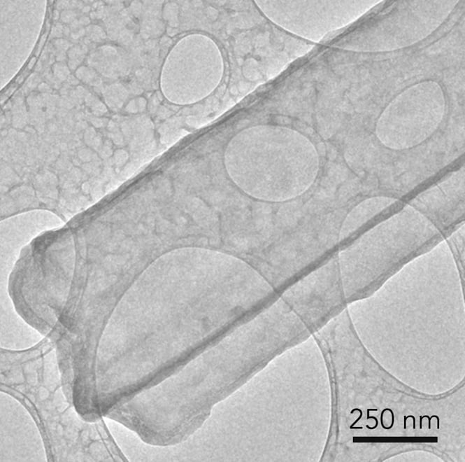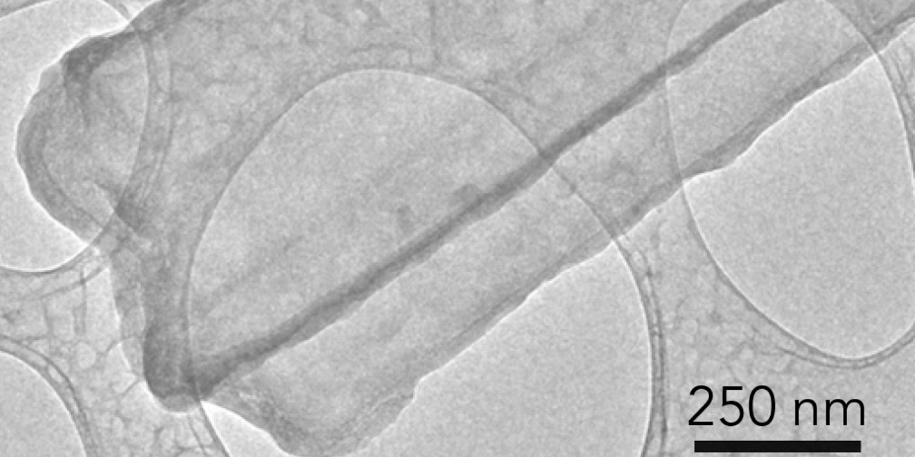
Researchers from Stanford and SLAC have made a new breakthrough in battery science that could help them to develop longer lasting batteries in the future. The team captured the first atomic-level images of dendrites, finger-like growth structures that form while batteries charge and hinder the ability of batteries to retain energy.
Dendritic growths have long posed a challenge for developers of high energy batteries, as they can puncture the barrier between battery compartments and cause short circuits or fires. The research group’s work used a technique called cryo-electron microscopy (cryo-EM), which won the 2017 Nobel Prize in Chemistry, to capture these new high-resolution images.
Materials science and engineering Professor Yi Cui, who led the group of Stanford Institute for Materials and Energy Sciences researchers behind the breakthrough, said that the group’s work successfully applied the imaging technology to a difficult but fruitful area — capturing previously elusive images of unstable metals.
“With cryo-EM, you can look at a material that’s fragile and chemically unstable and you can preserve its pristine state — what it looks like in a real battery — and look at it under high resolution,” said Cui in an interview with SLAC Media. “The lithium metal we studied here is just one example, but it’s an exciting and very challenging one.”
Before cryo-EM, scientists struggled to examine battery parts with transmission electron microscopy (TEM). According to Yuzhang Li, a fifth-year materials science and engineering Ph.D. student who co-authored the research, TEM damaged many of the sensitive battery materials, such as lithium metal.
“TEM sample preparation is carried out in air, but lithium metal corrodes very quickly in air,” Li said. “Every time we tried to view lithium metal at high magnification with an electron microscope the electrons would drill holes in the dendrite or even melt it altogether.”
Published in “Science,” the new cryo-EM images reveal a different rendering of the dendrites than past microscopic shots. They show that the lithium metal dendrite is a long, six-sided crystal, unlike the irregular, pitted shape shown in the TEM images. This level of detail allows scientists to understand how batteries work at the atomic level and why high-energy batteries found in cell phones, laptops and electric cars sometimes fail.
The researchers used a cryo-EM instrument at Stanford School of Medicine to analyze thousands of lithium metal dendrites exposed to different electrolytes (the medium that allows the flow of electrical charge through the battery). The group also looked at the solid electrolyte interphase, a coating that forms as the dendrite reacts with the surrounding electrolyte. As a battery charges and discharges, the coating forms on metal electrodes, potentially shortening battery life. Yanbin Li, a Ph.D. student in SIMES, said that controlling its growth is crucial in ensuring the battery runs efficiently.
Other strategies the researchers used to prevent damage from dendrites included adding chemicals to the electrolyte and creating a “smart” battery that shuts off automatically when it senses dendritic growths in the battery’s chamber. Moving forward, the researchers at Cui’s lab plan to concentrate on exploring the chemistry and structure of the SEI coating.
“We were really excited: This was the first time we were able to get such detailed images of a dendrite, and we also saw the nanostructure of the SEI layer for the first time,” Li said. “This tool can help us understand what different electrolytes do and why certain ones work better than others.”
Contact Sterling Alic at salic ‘at’ stanford.edu.
