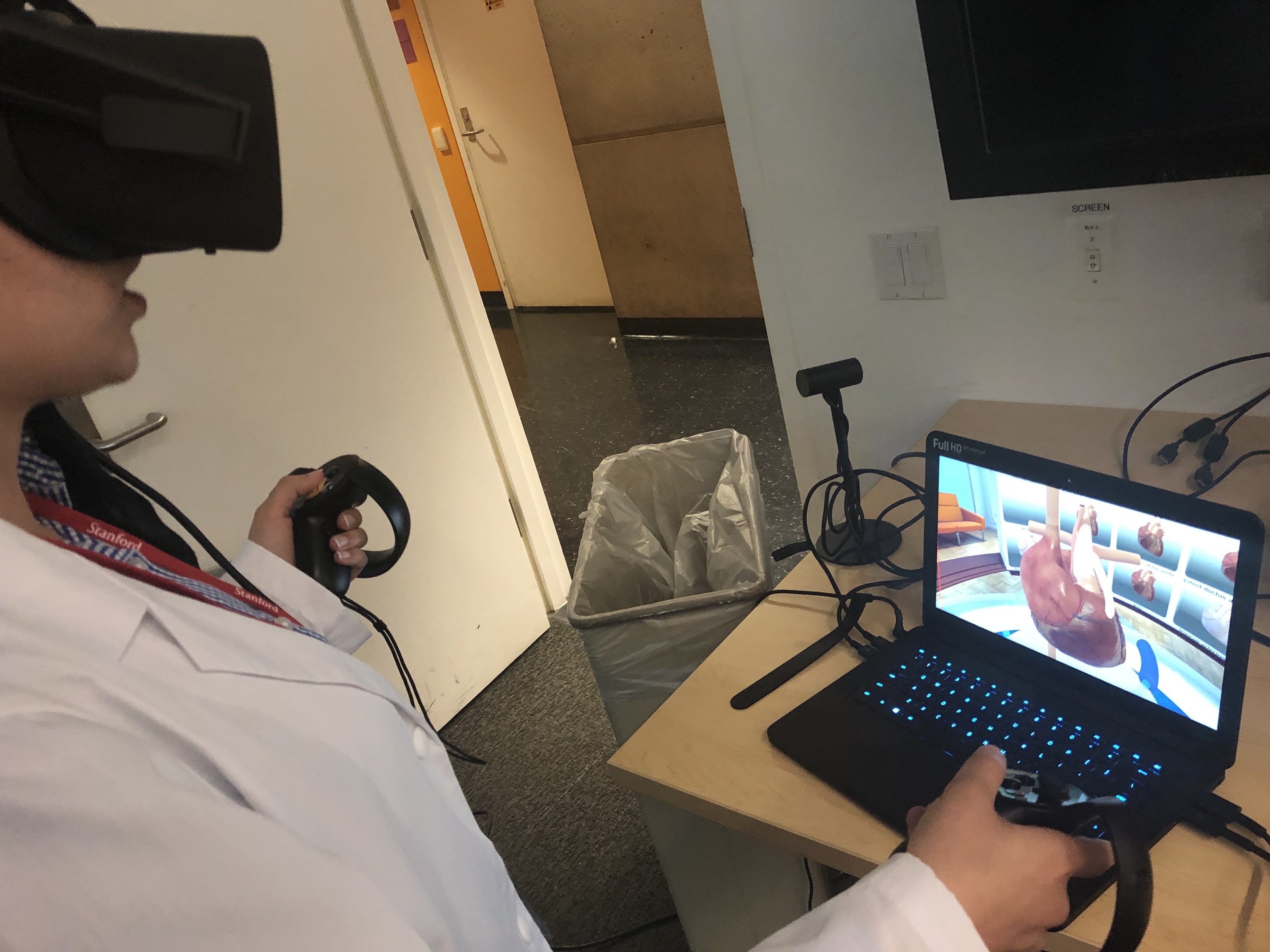
At the Lucile Packard Children’s Hospital, Stanford Medicine pediatric cardiologist David Axelrod is working on using virtual reality (VR) to model heart defects. The models serve both as educational tools for both learning surgeons and patients and their families.
Currently, the VR application has more than 15 heart models, which are used for instruction of heart anatomy and congenital heart defects. They are in use at 22 medical centers around the world, such as The University of Michigan.
“We built the hearts as prototypical congenital heart models based on a number of different patients, but each one is like a classical model of whatever heart defect it is depicting,” Axelrod said.
At programs like Stanford’s Pediatric Cardiology Fellowship Bootcamp, the VR hearts have been used to teach and train pediatric cardiologists. The VR models have also seen limited use at the School of Medicine, though Axelrod hopes to expand their integration into the curriculum and expand their educational potential in general.
“Right now what we really want to do is continue to build up the educational pedagogy behind [the application] so that it’s not just a model, but a full educational experience,” Axelrod said.
Recently, the team developed a camera application that lets users take, save and export photos of pre-surgical and post-surgical operation on the heart. Axelrod envisions that further educational features will be developed within the next nine months.
“If you’re able to train medical trainees more effectively on procedures that involve risk without actually having any real risk to a patient, then you’re going to get better outcomes in all areas in healthcare.” said David Sarno, founder of VR firm Lighthaus Inc. Axelrod partnered with Lighthaus to develop the models.
According to Sarno, the project started five years ago when Lighthaus Inc. built an application to visualize children’s congenital heart diseases for their parents. The models are built using the Autodesk Maya computer application and then processed through virtual reality programming application, Unity.
“It will be really beneficial for people to visualize their condition because humans are visually-inclined learners,” Sarno said. “The application will further make it easier for patients to retain knowledge about their problem and feel comfortable with the treatment.”
Axelrod also spoke about how the Lighthaus’s models helped parents of children with heart diseases visualize their child’s heart condition.
“It was just really neat to see the parents using the iPad application,” Axelrod said. “What used to be drawn out in pen and paper or even just flat pictures, [Lighthaus Inc.] has made more interactive and immersive so that families really understood the heart defects.”
The sister of a patient who was introduced to the VR heart model said that the model helped him better understand his family member’s condition and calmed his nerves.
“I always thought of my brother’s congenital heart defect, Hypoplastic Left Heart Syndrome (HLHS) as only something to be feared like a vague, dark looming figure that seemed too complex to understand,” she said. “I felt a sense of relief after using the Stanford Virtual Heart technology to better understand HLHS … It’s just something else to be able to use your own two hands and see for your own eyes what was once scary.”
She also believes the technology will be useful for family education.
“Without clear understanding, there is added stress that comes along because it can seem as though anything can happen without really knowing why,” she said. “The VR technology has the ability to close those gaps of confusion by providing reassurance and a sense of appreciation to clearly understand your loved one’s condition.”
Axelrod and his team are applying for a grant from the National Science Foundation. If it’s granted, it will allow the team to acquire a more sophisticated modelling technology. In the future, Axelrod wants to be able to use the models clinically.
“It is really hard to merge the world of what’s really important for medicine and what you want to develop from an educational standpoint with the inherent limitations of what you can and cannot build with the current VR experience,” Axelrod said. “I would love to use the application in the next two to five years for surgical planning, but then again the tech isn’t to the adequate level yet.”
“The technology does need to be at a whole new level,” Sarno said. “A simulation needs to have pressure, resistance, blood, so that the surgeon can actually feel like [they are] cutting into somebody.”
Although Axelrod and his team do not currently use patients’ heart scans because of poor resolution, they are in the process of improving the technology in hopes of eventually incorporating patient scans into the application.
Axelrod believes that VR technology will become increasingly integrated into medical centers in the future. He cited the existence of VR education centers in Nebraska as well as Stanford’s Neurosurgical Simulation and Virtual Reality Center.
“We are already beginning to see many medical centers using virtual, augmented, mixed reality applications for education and training,” Axelrod said. “I believe many more educators will see VR as a powerful tool in their curriculum and more programs will develop in the near future.”
A former journalist on the Knight Fellowship, Sarno initially investigated the use of interactive 3D graphics in journalism. When he founded Lighthaus Inc., the 3D graphic projects morphed into virtual reality.
“In healthcare, things stay relevant for a very long time, so it makes more sense to invest in the educational applications,” Sarno said.
The project has received funding from the Betty Irene Moore Children’s Heart Center and the Vice Provost for Teaching & Learning (VPTL) in addition to funding and hardware support from Oculus VR.
Contact Gokul Ramapriyan at gmramapriyan ‘at’ gmail.com.
